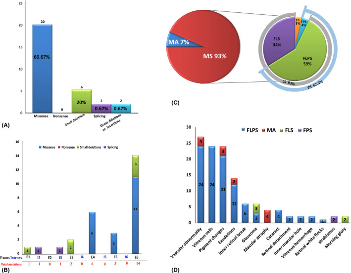
Fig. 1.

Overview of pathologic RS1 mutations identified in this study and complications of all the patients. (A) Number of RS1 mutations of different mutation types identified in this study. (B) Distribution of the RS1 variants identified in this study. Exons are numbered in black font, and introns are numbered in blue font. Total mutations are shown in red font. (C) Pie chart showing the structural changes in XLRS patients. Macular schisis was seen in 93% of patients, with lamellar schisis (LS) accounting for 93% and peripheral schisis (PS) accounting for 62.5%. (D) Distribution of the different complications observed in different types of patients. FLPS = complex type patients who had foveal, lamellar and peripheral schisis., FLS = foveo‐lamellar patients who had foveal schisis and lamellar schisis, but not peripheral schisis, FPS = foveo‐peripheral type patients who had foveal and peripheral schisis without lamellar schisis, FS = foveal schisis, MA = macular atrophy.
