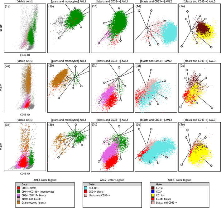
FIGURE 5.

Characteristics features of NPM1+ AML cases on radar plots: The distinctive features include low SSC, monocytic differentiation or prominent monocyte component, complete or partial expression of HLA‐DR, CD11c and CD15 on blast population. Three different patterns of NPM1+ AML are shown. The upper row shows a case with purely monocytic differentiation (1a–1c) CD11b+/CD14+ monocytic cells (seen as green dots) on SSC/CD45 and in two AML1 panel RPs. Only minimal CD34+ and CD117+ populations are seen (red and bright pink dots). (1d) HLA‐DR expression on leukemic cells (cyan dots). (1e) Leukemic cells express CD15 (maroon dots) and CD11c+ (yellow dots). The middle row shows a case of blastic NPM1+ AML with almost no monocytic component: (2a) Low SSC/CD45dim blasts merging with maturing granulocytes. (2b) Granulocytes (brown dots) with minimal monocyte population (green dots). (2c) CD117 and partial CD34 expression (bright pink and red clusters). (2d) Only partial HLA‐DR expression in blast population (cyan dots). (2e) Low CD15 expression (maroon dots) and partial CD11c expression (yellow dots). The lower row shows a case of myelomonocytic NPM1+ AML: (3a–c) Low SSC, CD117+ and partly CD34+ blasts (pink/red dots) and a distinct monocytic population (green dots). (3d) Uniform expression of HLA‐DR (the cyan cluster). (3e) Uniform expression of CD11c (the yellow cluster but no CD15 or CD2 expression). Axes of RPs are arranged as shown in Figure 2. Color legends are arranged based on descending color precedence in each panel with intent to highlight distinctive markers. AML, acute myeloid leukemia; RP, radar plot [Color figure can be viewed at wileyonlinelibrary.com]
