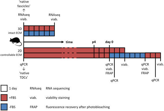
Figure 1.

Schematic overview of experimental setup. Complete fascicles were cultured for 6 days in serum‐rich (+FBS) or serum‐free (−FBS) medium. mRNA was sequenced, cell viability was determined, and both readouts were compared to freshly isolated fascicles (“native fascicles”). TDCs were expanded in +FBS medium, and at sub‐confluency in passage 4 (day 0) medium was switched to +FBS or −FBS medium. Gene expression, cell viability, and functional intercellular communication were monitored over time, and gene expression was compared to TDCs directly after fascicle digestion (“native TDCs”). 2D, two‐dimensional; ECM, extracellular matrix; FBS, fetal bovine serum; mRNA, messenger RNA; qPCR, quantitative polymerase chain reaction; TDCs, tendon‐derived cells [Color figure can be viewed at wileyonlinelibrary.com]
