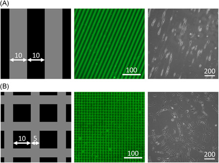
Figure 2.

TDC morphology and orientation were controlled using microcontact printed collagen I patterns. Schematic of aligned (A) and random (B) microcontact printing patterns (1), with collagen I printed surfaces in gray and Pluronic‐F127–coated surfaces in black. Microcontact printed substrates with collagen I fluorescently stained in green (2) and resulting phase contrast microscopy images of seeded TDCs (3). All sizes are given in micrometers. TDCs, tendon‐derived cells [Color figure can be viewed at wileyonlinelibrary.com]
