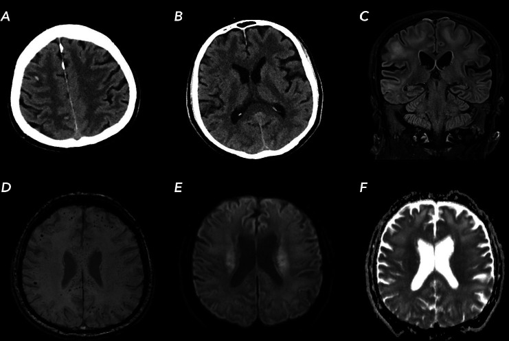
Figure 1.
Brain CT and MR images of our patient with COVID-19 infection. (A, B) axial CT images showing subcortical small haemorrhages and symmetrical hypodense areas in the supratentorial white matter. (C) MR turbo inversion recovery magnitude T2-weighted image showing subcortical T2-hyperintensies in the subcortical areas. (D) MR susceptibility weighted images showing widespread microbleeds in white matter and splenium. (E, F) paired diffusion-weighted imaging and apparent diffusion coefficient showing restricted diffusion in the corona radiata.

