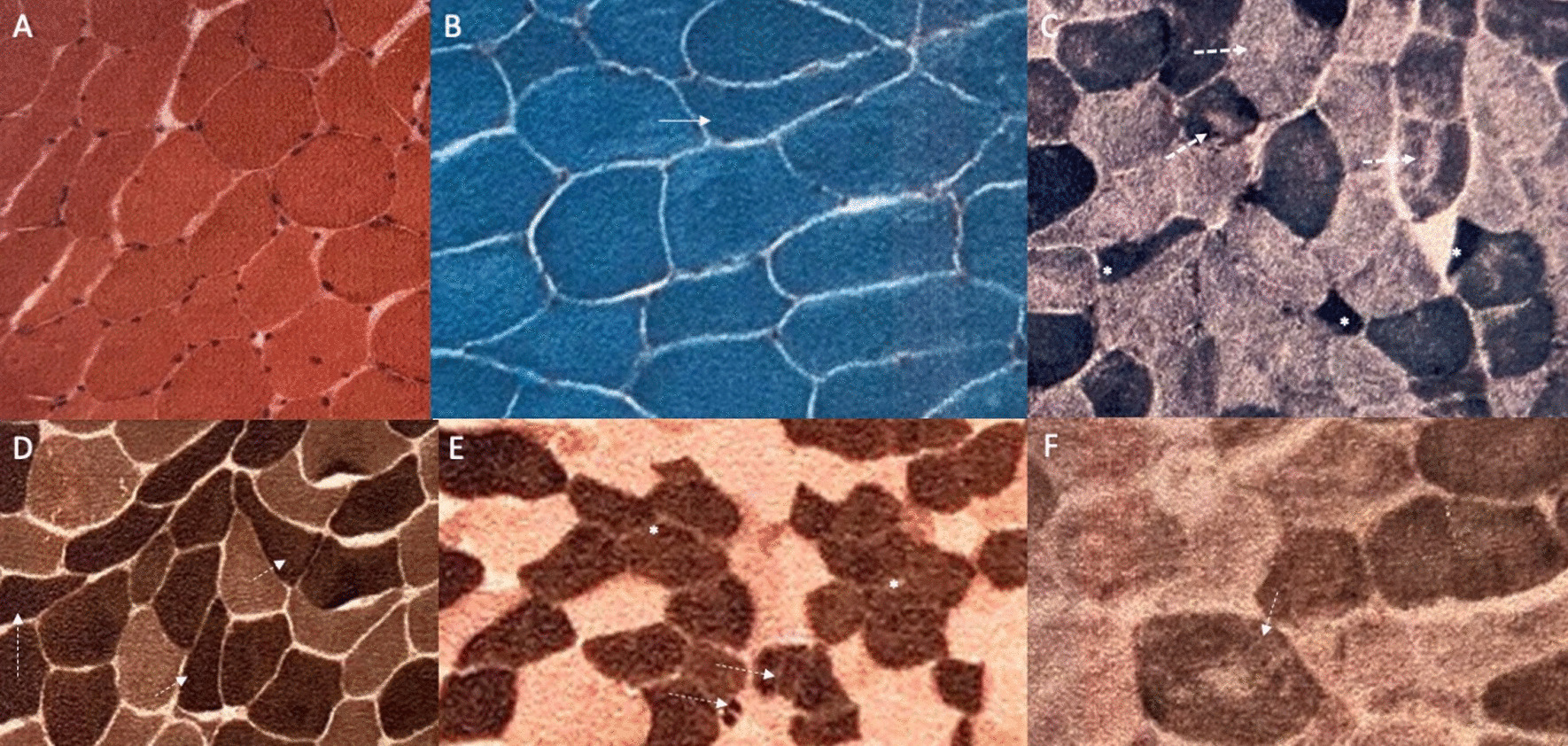
Fig. 5.

Muscle biopsy features. a Hematoxylin–eosin (HE) staining disclosing mild variation in muscle fiber diameter and nuclei centralization; b modified Gömöri trichrome staining showed angular fibers (white arrow); c NADH-tetrazolium reductase (NADH-TR) showed target fibers (white arrows) and angular fibers (asterisk); d ATPase stain at pH 4.6 and e ATPase stain at pH 9.4 showed angular fibers (white arrows) and tendency to type grouping formation (asterisks); f Cytochrome C-oxidase (COX) reaction disclosed focal central areas with reduced staining
