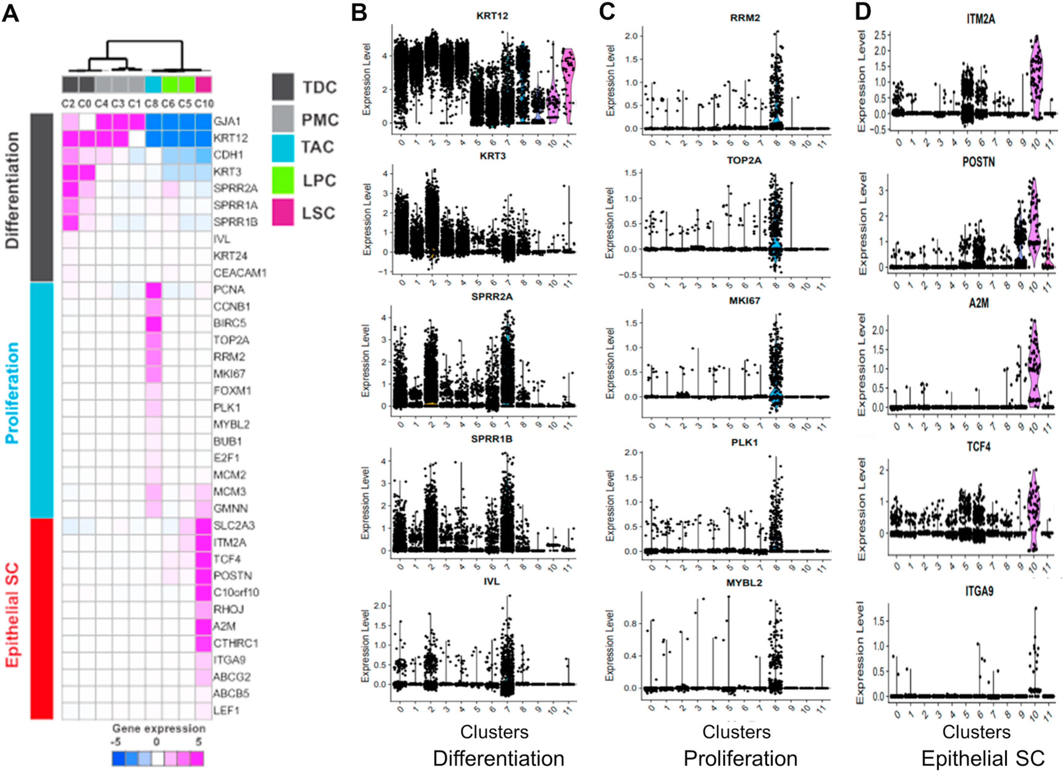
Fig. 3.

Identification of the subtypes of limbal cells. Known markers (n = 36) classified based on limbal cell differentiation status: Differentiation, proliferation, and epithelial stem cell. A. Unsupervised clustering of the expression profile of 36 known markers. Expression level was median centered of z scaled expression. B-D. Violin plot shows marker genes of differentiation, proliferation, and epithelial stem cells respectively.
