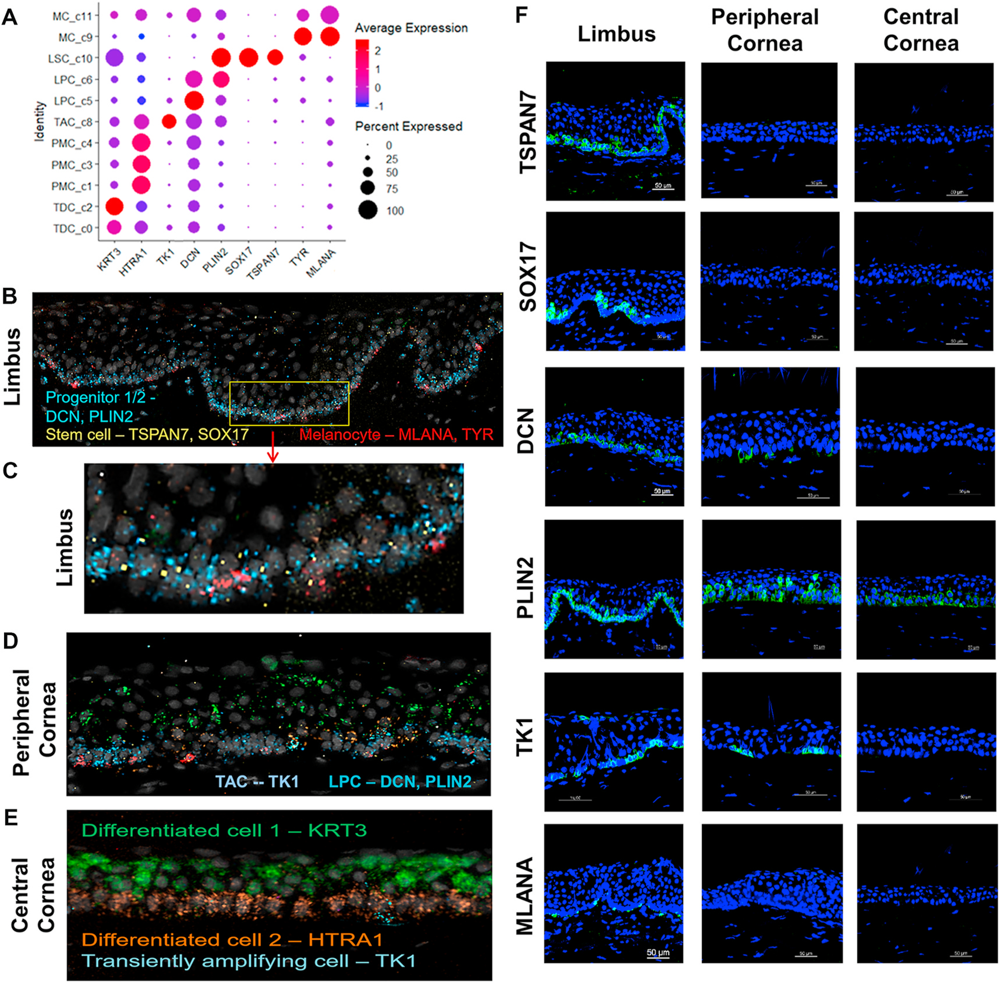
Fig. 6.

Validation of cell type-specific marker genes in five sub cell types at transcript and protein levels using RNAscope and immunofluorescent staining. A. Dot plot shows the expression of 9 marker genes selected from five cell-types. B-E. RNAscope in-situ validation shows the spatial expression of the transcripts of cell type-specific markers in three different regions of corneal tissues. Progenitor cells on limbus were identified by DCN and PLIN2 in blue, Stem cells by TSPAN7 and SOX17 in yellow, and Melanocytes by MLANA and TYR in red (B,C). On peripheral cornea more cell types were observed by known markers such as TK1 for TAC, DCN and PLIN2 for LPC and TDC for KRT3 (D). On central cornea, KRT3 for TDC and HTRA1 for PMC were expressed (E). F. Immunofluorescent staining shows the spatial localization of these marker proteins at limbus, peripheral, and central cornea.
