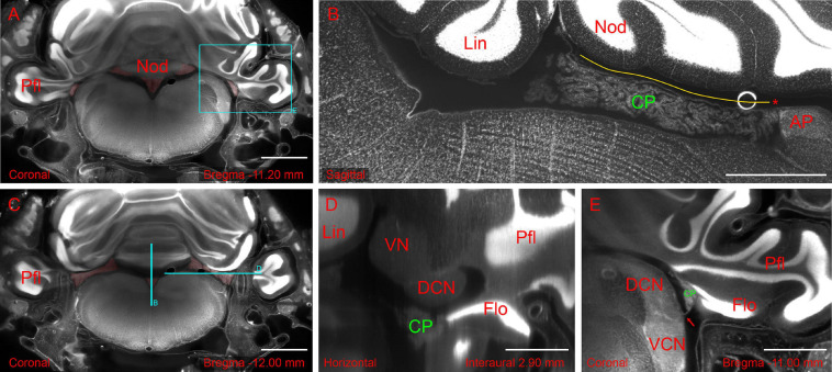
FIGURE 2.
Hindbrain slices from light-sheet 3D dataset of the whole-mount brain-bone preparation showing the extent and association of the CP. (A) Coronal slice showing CP attachment (highlighted in red) at the nodulus and paraflocculus. Scale bar: 2 mm. (B) Sagittal slice (different animal from A) showing the CSS with overlying tela coroidea (yellow line) and attachment to area postrema (asterisk). Scale bar: 1 mm. (C) Coronal slice showing the MHS (highlighted in red). Same animal as in (A). Scale bar: 2 mm. (D) Horizontal slice detail (reslice of stack in A,C) showing the dorsal cochlear nucleus (DCN) and flocculus (FL). Scale bar: 1 mm. (E) Coronal slice detail from animal in (A) showing the foramen of Luschka (arrow) with the associated CP. Scale bar: 1 mm. All sections were stained with TOPRO for cell nuclei. Autofluorescence signal is also visible. Cyan rectangle on panel (A) and cyan lines on panel (C) indicate x, y positions of panels (B,D,E).

