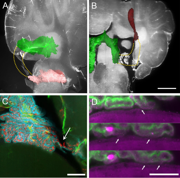
FIGURE 4.
MHS-LHS junction after removal of temporal bone. (A) Overlay of plexus vascular segmentation with a substack of unsegmented hindbrain. Red, LHS; green, SS and MHS. The stippled area in yellow corresponds to the location of the missing part of the CP. Sagittal view. (B) Similar to (A), resliced to horizontal plane. Scale bar: 1 mm. (C) COLM image of a contact region between CP and DCN in a sample where temporal bone has been removed. The plexus appears ripped (arrow) but contact with DCN is not removed. Scale bar: 200 μm. (D) Magnification of the CP-DCN contact (same animal as in C) showing the indentation marks left by CP villi on DCN ependymal surface. In some positions (arrows) the CP appears connected to the DCN surface. Scale bar: 100 μm.

