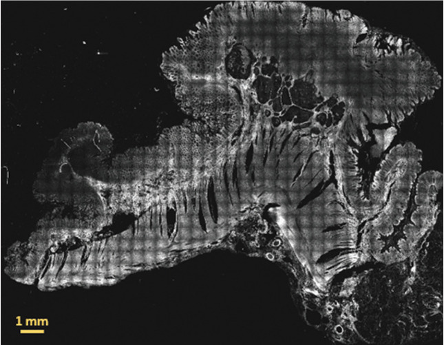
Figure 3.
Two-photon fluorescence image of a whole 30 μm thick paraffin-embedded tissue slide with sample 86 diagnosed as low grade adenocarcinoma. The signal originates mainly from mitochondrial NADH in the cell cytoplasm and from elastic fibers and other fluorescent molecules in the extracellular matrix. This image has been obtained by merging 37 by 29 image tiles, resulting in an overall field of view: 18.907 mm by 14.819 mm

