Figure 5.
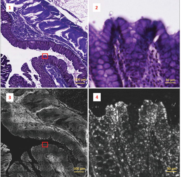
Two-photon fluorescence image (bottom-left) acquired from a 30 μm thick paraffin-embedded tissue slide with sample 69-2 diagnosed as tubular adenoma with low grade dysplasia and its co-registered corresponding H&E image (top-left). Two-photon fluorescence image was obtained by concatenating 9 by 12 image tiles. The overall field of view results in 4.599 mm by 6.132 mm. Detail marked by the red box in the images on the left is represented on a magnified scale on the right. Colonic crypts and goblet cells can be identified in the H&E crop (top-right), but these features are not appreciable on the corresponding two-photon fluorescence crop (bottom-right)

