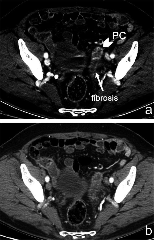
Figure 3.

a, b Peritoneal recurrence in a 54-year-old man with diffuse GC, who underwent cytoreductive surgery and HIPEC. Monoenergetic images at 40 keV (a) show a conspicuous vascularization of a nodule of PC (arrowhead) in the left external iliac site, next to an area of fibrosis (arrow) and without ascites. It is not possible to distinguish between PC and fibrosis at standard 140 kVp images (b)
