
FIG. 2.
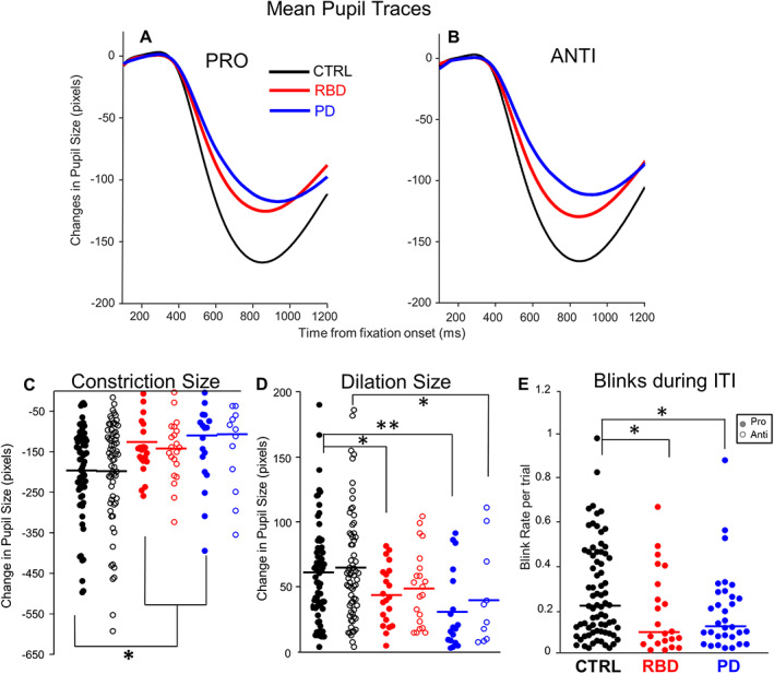
Mean pupil traces for each patient group are represented during the pro‐ (A) and anti‐ (B) saccade conditions over time. (C) Constriction size was defined as the pupil size at the greatest constriction after fixation appearance. (D) Dilation magnitude was defined as the pupil size at stimulus onset minus the pupil size at the time of greatest constriction during FI, reflecting the increase of pupil size after constriction. (E) Median blink rate during the inter‐trial interval (ITI). *P ≤ 0.05, **P ≤ 0.01, ***P ≤ 0.001. CTRL, healthy, age‐matched controls; RBD, rapid eye movement sleep behavior disorder; PD, Parkinson's disease. [Color figure can be viewed at wileyonlinelibrary.com]