
FIGURE 3.
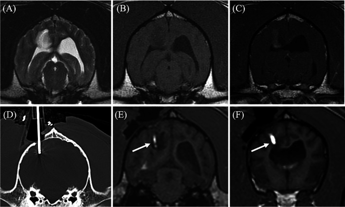
Transverse diagnostic imaging of a live dog. (A) T2 weighted transverse image. A hyperintense mass is present within the right parietal lobe. (B) The mass appears hypointense on T1 weighted (T1W) images and has moderate heterogeneous contrast enhancement (C). (D) Transverse computed tomography (CT) set on a bone window showing the placement of the biopsy needle within the mass. (E) Transverse 3D T1W image during convection enhanced delivery (CED) of molecularly targeted therapeutics into the mass. The arrow is displaying the therapeutics which is infused with gadolinium making the needle placement and trajectory visible on magnetic resonance imaging. (F) A more rostral transverse 3D T1W image compared to (E) where a separate needle was placed. The CED guide allowed for multiple needles to be placed within the mass for a more even distribution of the therapeutics