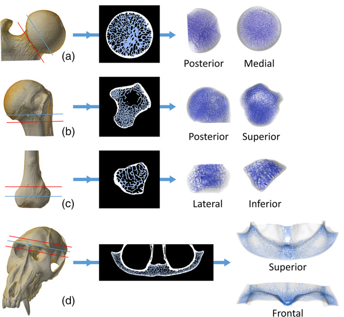
FIGURE 4.

Semi‐automatic isolation of cancellous bone in the femoral head of Symphalangus syndactylus (a), the proximal humerus of Alouatta caraya (b), the distal fibula of Cercopithecus albogularis (c) and the brow ridge of Mandrillus sphynx (d). The 3D μCT scan is cut (red line) to limit the cancellous isolation to a region of interest. The results are here shown on a single 2D slice (indicated by the blue line on the 3D scan) and on the full 3D μCT stack (the cutting planes used to isolate the 3D regions of interest is shown in red)
