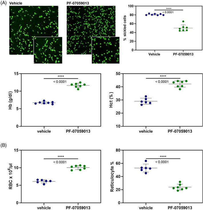
FIGURE 1.

(A) Left: Representative images of red blood cells from vehicle treated and PFE‐07059013 treated animals, following 4 h of hypoxic exposure. Inset: Expansion of section of depicted field to highlight differences in number of sickled cells. Right: Individual values for RBC sickling for vehicle treated and PF‐07059013 treated mice. The treated animals showed a robust and statistically significant reduction in RBC sickling. (B) Markers of hemolytic anemia measured for vehicle treated (blue) and PF‐07059013 treated (green) mice. Note, PF‐07059013 treated animals showed increases in RBC count, hemoglobin (Hb), and hematocrit (Hct), indicating a substantial reduction in hemolysis. PF‐07059013 treated animals also showed a significant decrease in reticulocytes, suggesting decreased hematopoiesis in the PF‐07059013 animals, due to the decreased hemolysis
