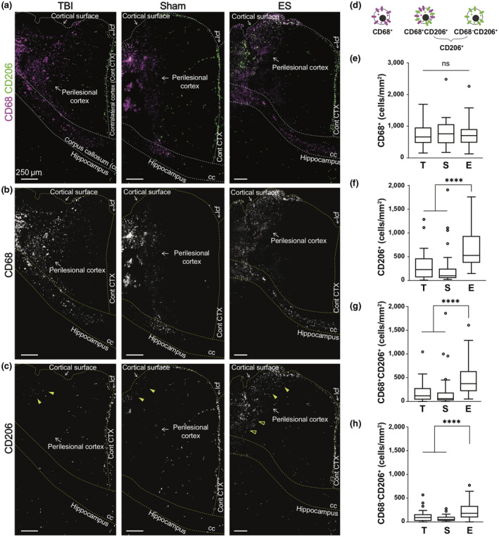
FIGURE 2.

Effect of electrical stimulation (ES) treatment on CD68 and CD206 expression in perilesional cortex at 7 days post‐traumatic brain injury (TBI). (a) Representative low magnification images of CD68 (magenta) and CD206 (green) staining from the ipsilateral cortex to the hippocampus. (b) CD68+ cells mostly appeared at the perilesional cortex and corpus callosum (cc), but not in the hippocampus. CD68+ cells were shown to be distributed similarly in all experimental groups. (c) CD206+ cells appeared relatively less than CD68, and they were observed at the longitudinal cerebral fissure (lcf), ipsilateral cortex, and hippocampus. Within the perilesional cortex, CD206+ cells were observed near the cortical surfaces in all experimental groups (filled arrowheads). On the other hand, CD206+ cells at the deeper cortex were only observed in the ES group (empty arrowheads). (d) Illustration of three subtypes of immune cells based on CD68 and CD206 expression. (e–h) The number of subtype cells across the entire perilesional cortex. T, S, and E on the x‐axis represent untreated TBI, sham and ES groups. (e) The number of CD68+ monocyte/macrophages/microglia did not exhibit a significant difference by treatment. In the entire perilesional cortex, the number of (f) CD206+ cells, (g) CD68+CD206+ cells, and (h) CD68−CD206+ cells significantly increased after ES treatment. Data are represented as mean ± SD. N = 4, 3, 6 animals with two tissue slices analyzed for untreated TBI, sham, ES; ****p < 0.0001, Kruskal–Wallis one‐way ANOVA with Dunn's post hoc analysis
