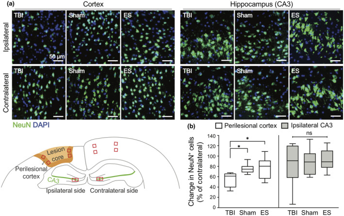
FIGURE 7.

Effect of treatment on neuronal viability in the ipsilateral cortex and hippocampus at seven days post‐traumatic brain injury (TBI). (a) Representative fluorescence images of NeuN+ neurons (green) in the cortex (left three columns) and CA3 in the hippocampus (right three columns). The images in the first row and second row represent the NeuN in the ipsilateral and contralateral sides, respectively. (b) Quantitative analysis of the change in the number of NeuN+ cells in the ipsilateral ROI compared to the contralateral side. In the ipsilateral cortex, NeuN+ cells remained more in the sham and ES groups compared to untreated TBI group. On the other hand, the changes of NeuN+ cells in the ipsilateral CA3 region were not significantly different across the groups. For the analysis of NeuN+ cells, four ROIs were taken from each ipsilateral and contralateral cortices, and two ROIs were taken from each ipsilateral and contralateral CA3 regions. N = 4, 3, 6 animals with two coronal sections analyzed for untreated TBI, sham, ES; *p < 0.05, Kruskal–Wallis one‐way ANOVA with Dunn's post hoc analysis
