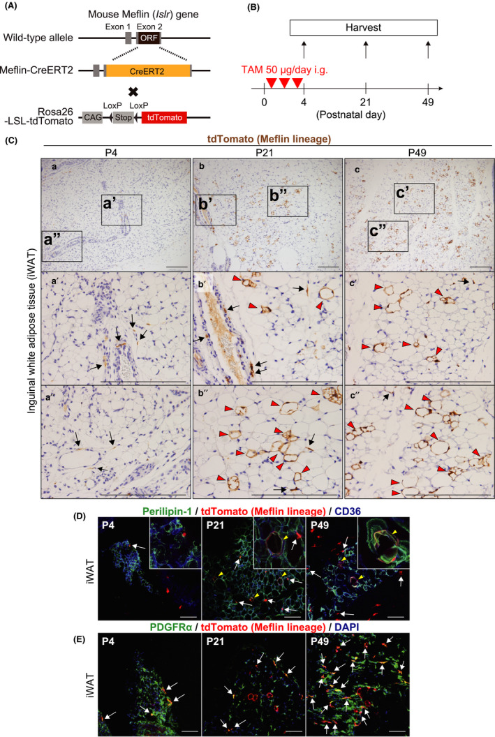
FIGURE 1.

Meflin+ cells are fibroblastoid cells that differentiate into white adipocytes in the postnatal period. (A) A diagram of the generation of Meflin‐CreERT2; LSL‐tdTomato mice. ORF; open reading frame, CAG; chicken ß‐actin promoter with cytomegalovirus enhancer and Stop; stop element. (B) The experimental design for lineage tracing of Meflin+ cells in the postnatal period. The Meflin‐CreERT2; LSL‐tdTomato pups were administered 50 µg tamoxifen (TAM) at P1, P2 and P3 to label Meflin+ cells with tdTomato. (C) iWAT harvested from Meflin‐CreERT2; LSL‐tdTomato mice was stained for tdTomato by IHC. tdTomato+ cells (brown) represent Meflin+ cells at P4 (a) and their descendants (Meflin lineage cells) at P21 (b) and P49 (c). Boxed regions in a–c are magnified in lower panels (a′–c′ and a″–c″). Arrows denote tdTomato+ stromal cells with a fibroblastoid morphology. Red arrowheads indicate tdTomato+ terminally differentiated mature white adipocytes (Meflin lineage cells). Scale bars, 200 µm. (D, E) iWAT harvested from Meflin‐CreERT2; LSL‐tdTomato mice was stained for the adipocyte marker Perilipin‐1 (D) and the fibroblast marker PDGFRα (E) by IF (green). CD36 is a marker of adipocytes and endothelial cells (blue). Magnified regions are indicated in insets. Arrows indicate tdTomato+ stromal cells with a fibroblastoid morphology, whereas arrowheads show tdTomato+ mature adipocytes. Note that tdTomato+/PDGFRα+ cells were observed throughout P4, P21 and P49 in (E). Scale bars, 100 µm
