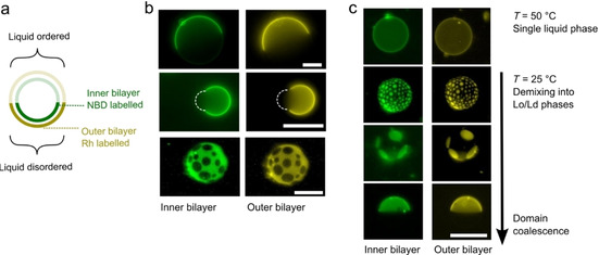
Figure 5.

Domain alignment in two‐layered vesicles. a) Schematic showing inter‐bilayer domain alignment in a two‐layered ternary mixture with vesicles exhibiting liquid‐liquid phase separation. b) Fluorescence images of inner and outer bilayers of two‐layered vesicles including spherical (top), bulging (middle), and multi‐domain vesicles (bottom). Dotted white lines represent bulging areas of the vesicle, not visible under fluorescence. Domains are aligned in all cases. c) Domains remain aligned through the demixing process, when vesicle were cooled below their miscibility temperature, from a fully mixed single liquid regime to a phase‐separated one. Vesicles were composed of DPhPC/EggSM/cholesterol 1 : 1 : 3. Scale bars: 20 μm.
