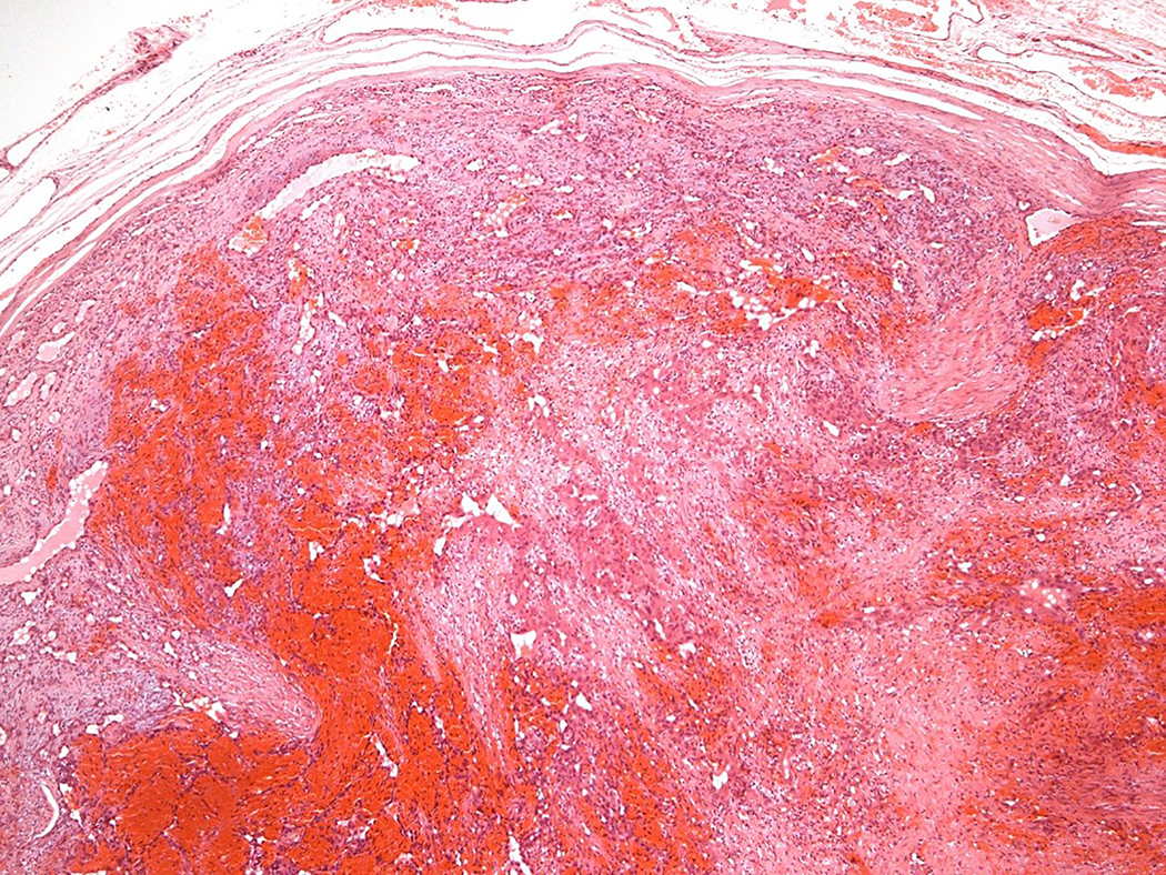
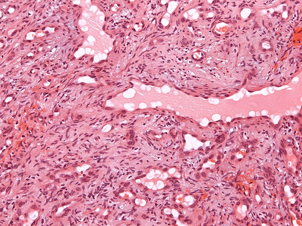
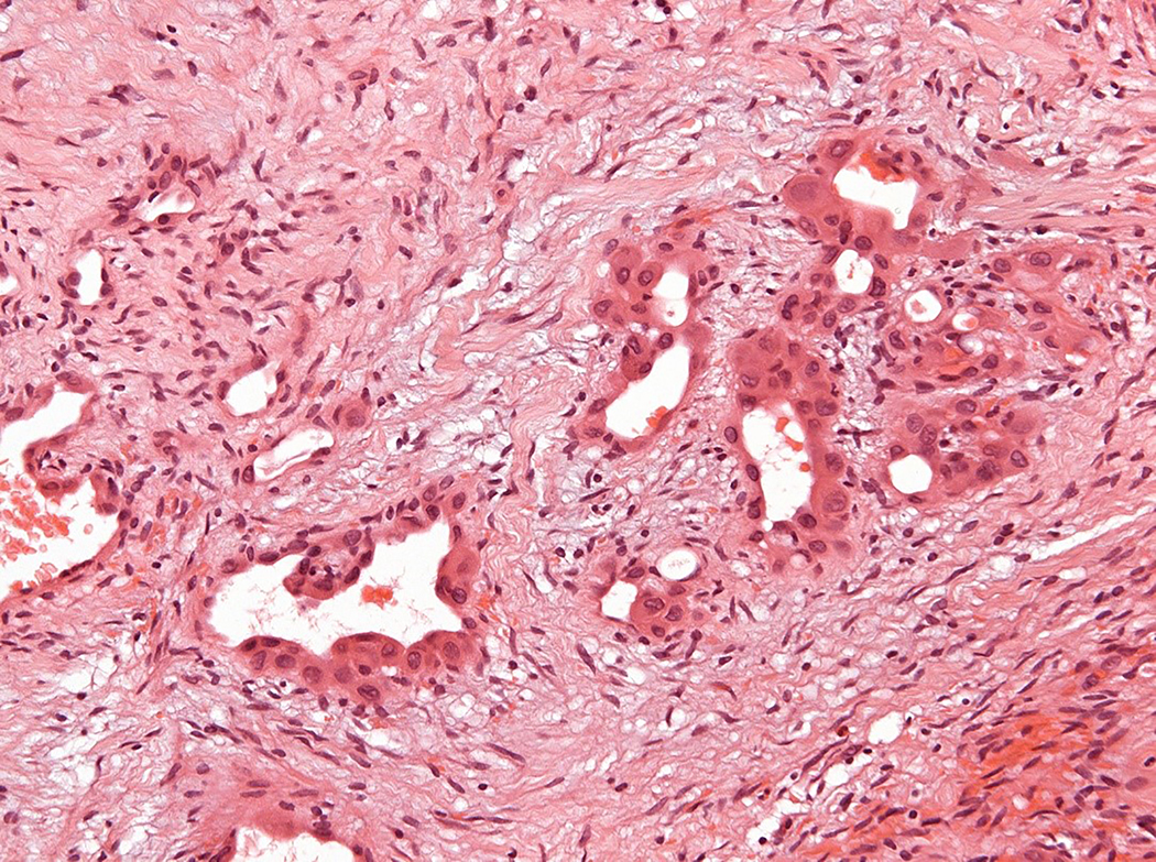
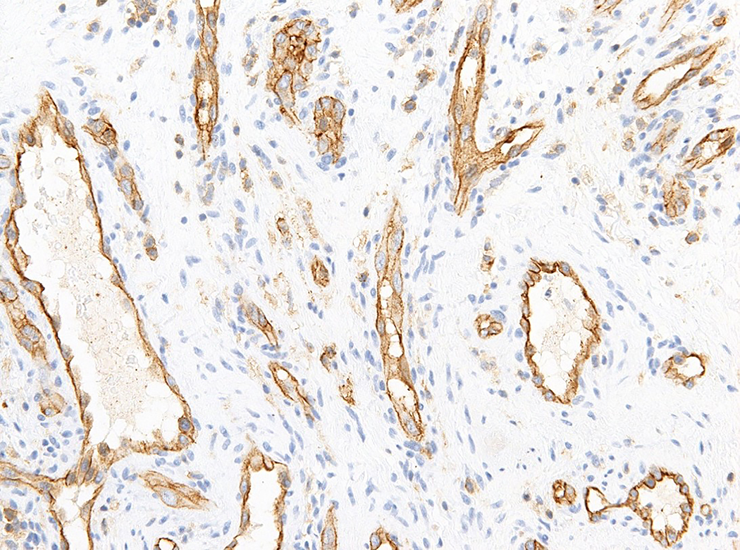
Figure 1.
Intravascular epithelioid hemangioma – capillary hemangioma type (case 12). A. the lumen of the vein is completely obliterated by vascular proliferation. B. Well-formed vascular channels are lined by a single layer of epithelioid endothelial cells. C. Mild nuclear atypia of endothelial cells is seen and there in no mitotic activity. D. Epithelioid endothelial cells are highlighted by CD31 immunohistochemistry.


