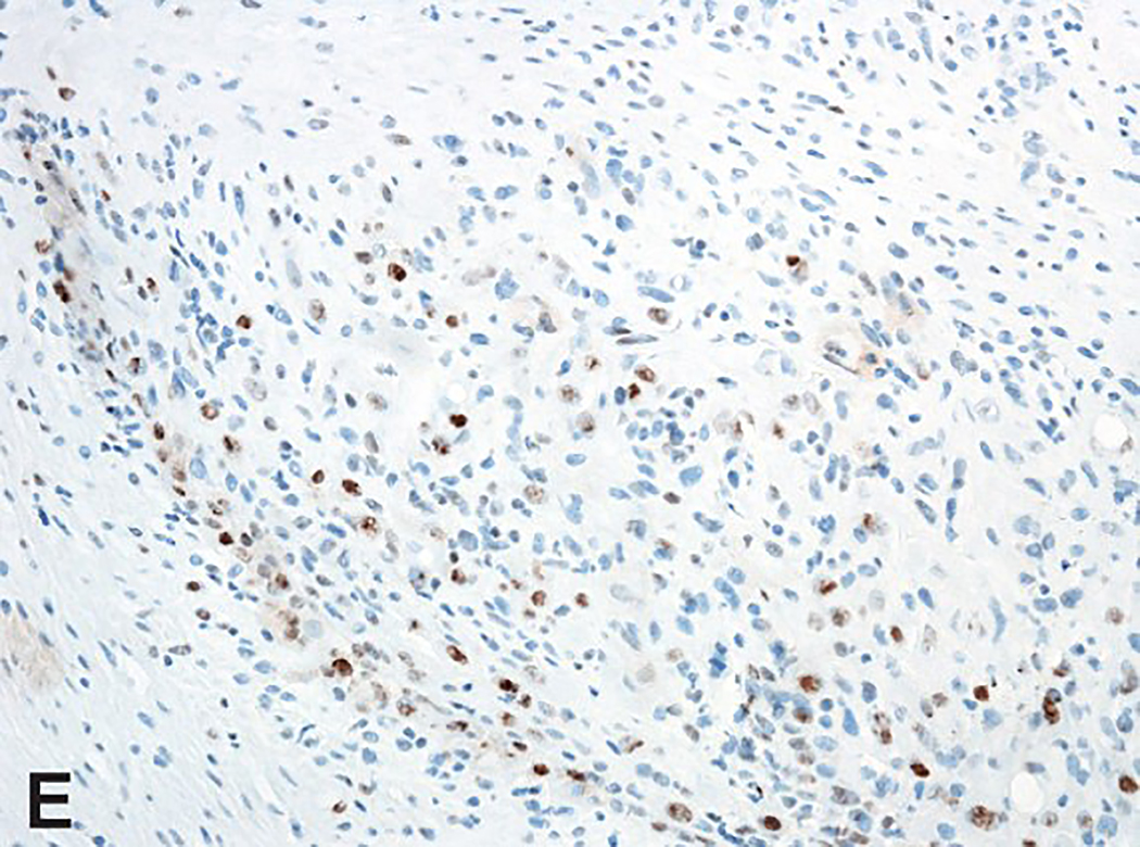
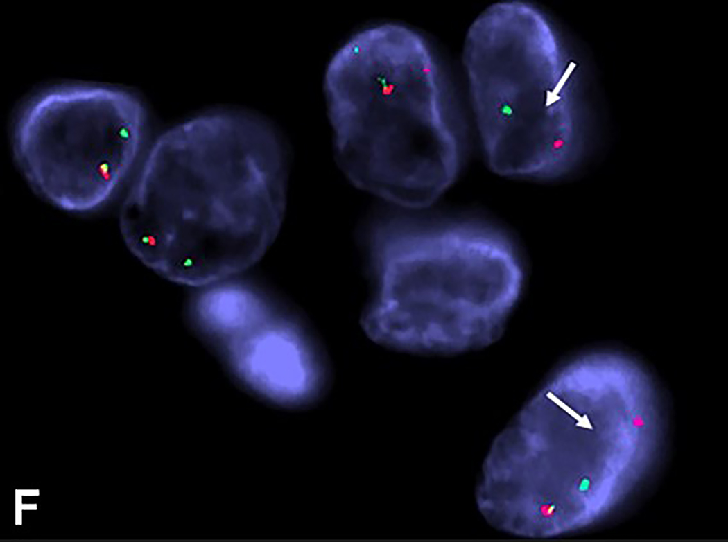
Figure 3.
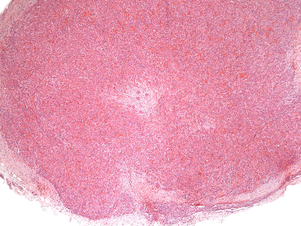
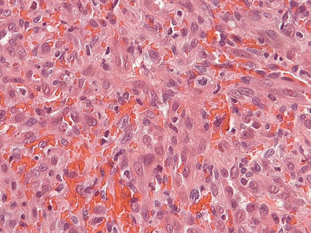
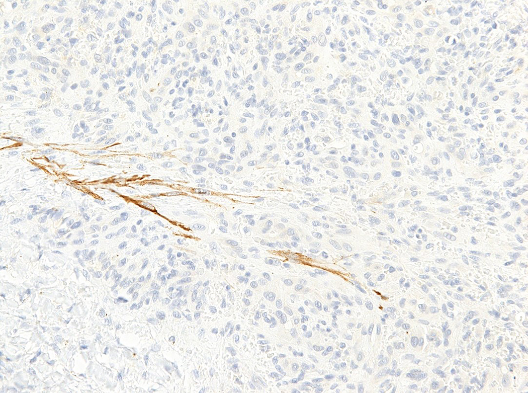
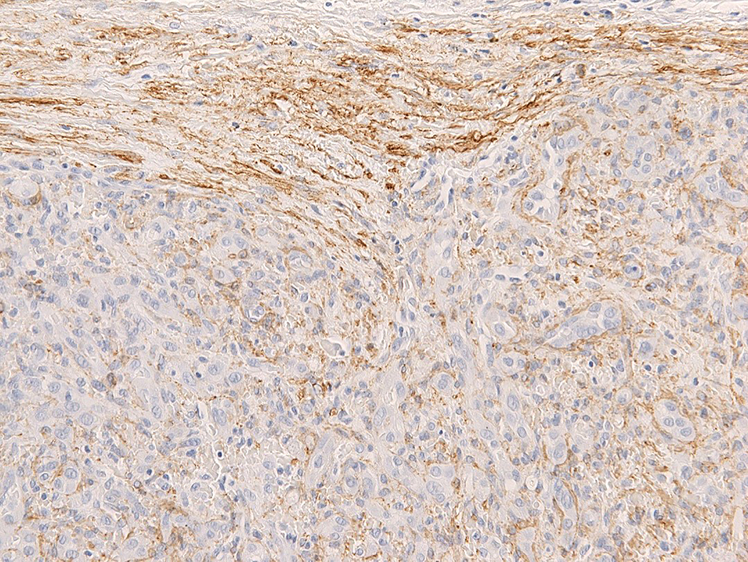
Intravascular epithelioid hemangioma – solid type (Case 6). A. Vascular differentiation of this intraluminal proliferation is not readily apparent at low-power magnification. B. Epithelioid endothelial cells display mild to moderate nuclear atypias. Note also brisk mitotic activity. C. Desmin immunohistochemistry can be discontinued or patchy due to vessel wall stretching. D. Immunohistochemistry for smooth muscle actin nicely demonstrated the wall of a distended vein. E. FOSB immunohistochemistry. About 30% of tumor cells display nuclear positivity for FOSB. Interestingly, this case lacked rearrangements of FOSB gene by FISH analysis. F. Case 6 was the only tumor that proved positive for FOS gene break-apart by FISH.

