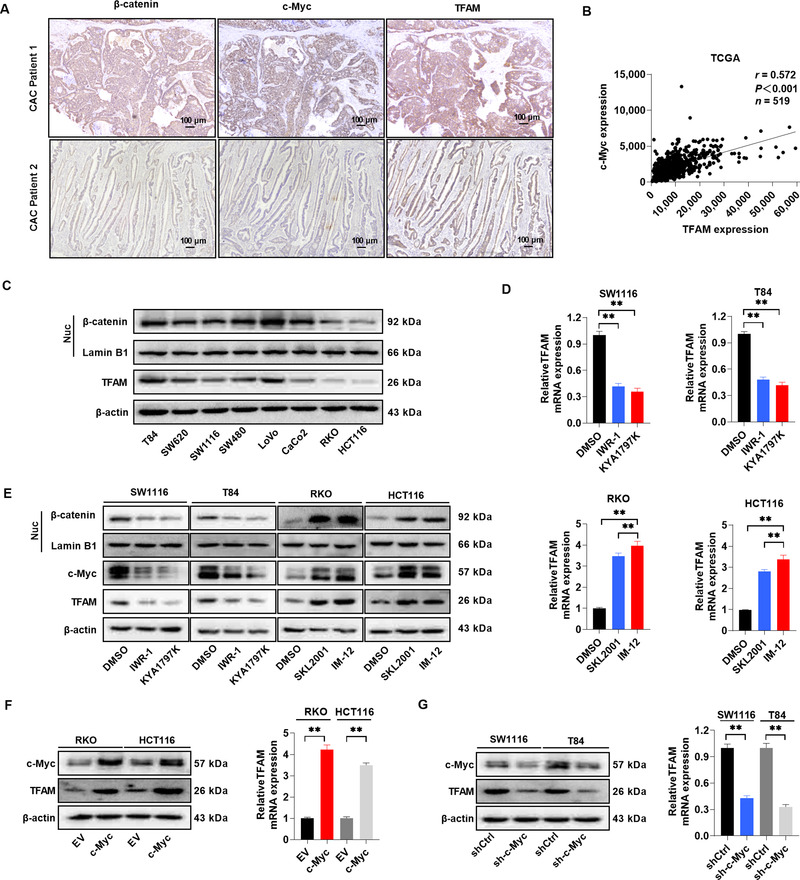
FIGURE 7.

β‐catenin induces upregulation of TFAM through c‐Myc in CRC cells. (A) Representative IHC staining images of β‐catenin, c‐Myc, and TFAM in CAC tissues from different patients. Scale bar, 100 μm. (B) Correlation between c‐Myc and TFAM mRNA expression in TCGA database. (C) WB for the expression of nuclear β‐catenin and TFAM expression in different CRC cell lines. Lamin B1 (a nuclear envelope marker) and β‐actin were used as loading controls in the nuclear and cytoplasmic fractions, respectively. (D) RT‐qPCR analysis for TFAM expression in CRC cells treated with Wnt/β‐catenin signaling pathway inhibitors (IWR‐1 and KYA1797K) or agonists (SKL2001 and IM‐12) as indicated. (E) WB for the expression of nuclear β‐catenin, c‐Myc, and TFAM in CRC cells treated as indicated. (F) WB and RT‐qPCR analysis for the expression of c‐Myc and TFAM in RKO and HCT116 cells after lentiviral transfection with EV or c‐Myc expression vector. (G) WB and RT‐qPCR analysis for the expression of c‐Myc and TFAM in SW1116 and T84 cells expressing shCtrl or shRNA against c‐Myc (sh‐c‐Myc). Error bars represent mean ± SD. Data were analyzed using Mann‐Whitney U test. *P < 0.05, **P < 0.01. Abbreviations: TFAM, mitochondrial transcription factor; TCGA, The Cancer Genome Atlas; EV, empty vector; SD, standard deviation
