
Figure 1.
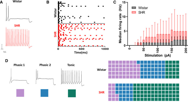
Stellate ganglia neurons of the spontaneously hypertensive rat have a hyperactive phenotype with an altered electrophysiological profile as observed by perforated patch-clamp recordings. A, Example traces showing the response of sympathetic neurons from control Wistar and prehypertensive SHRs (spontaneously hypertensive rats) to 150 pA of current injection for a duration of 1000 ms. B, Time course of induced firing following a 1000-ms 150-pA current injection in 30 example Wistar and SHR neurons. C, Median response of Wistar (black) and SHR (red) neurons to a range of current injections (Wistar, n=66; SHR, n=69; mixed-effects model: P<0.0001). D, Examples of sympathetic neuron firing rates are identified by their classical nomenclature. Here, the color scheme used to indicate these subtypes throughout the article is indicated by a colored square. A trend toward tonic firing neurons was observed in the SHR, shown as percentage of cells conforming to each subtype (Wistar, n=66; phasic 1, n=29; phasic 2, n=24; tonic, n=13; SHR, n=73; phasic 1, n=9; phasic 2, n=17; tonic, n=47; χ2=12.37, df=2, P=0.0021).