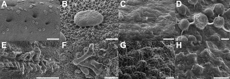
Figure 17.
Scanning electron micrographs of chorionic microstructures on the eggs A–DTrolicaphylliumsarrameaense comb. nov. E–HChitoniscus sensu stricto A, B mushroom-like granula C, D, G, H surface microsculpture C surface of the granula D exochorionic surface microstructures E, F pinnae. Scale bars: 100 µm (A, E), 20 µm (B, F), 10 µm (G), 5 µm (D), 3 µm (H), 1 µm (C).

