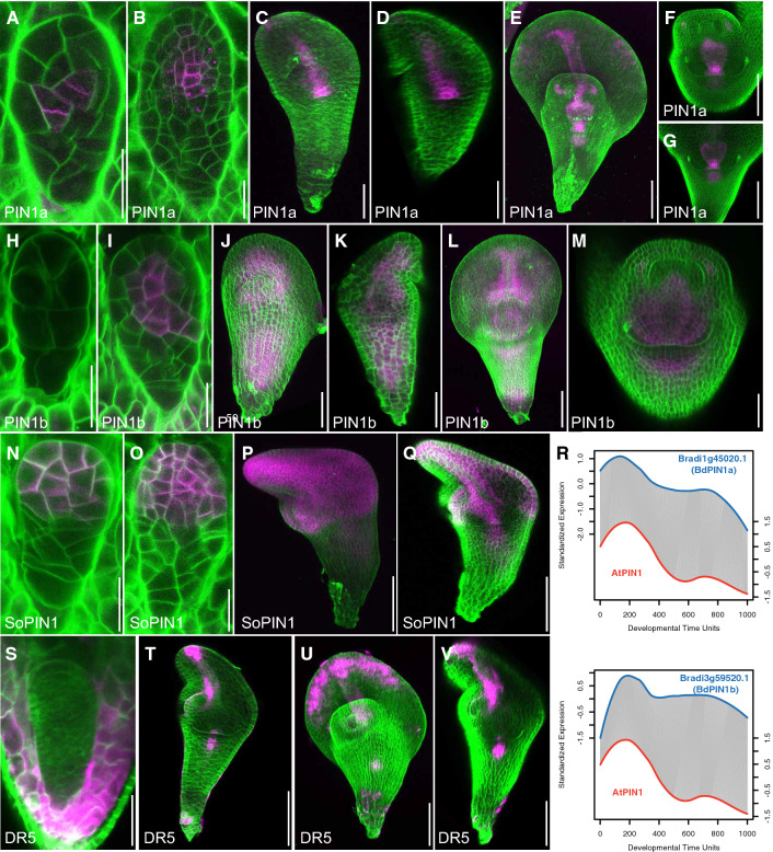
Fig. 7.
Auxin transport and response during Brachypodium embryogenesis. (A–Q) Localization of PIN1a-Citrine (A–G), PINb-Citrine (H–M) and SoPIN1-Citrine (N–Q) during Brachypodium embryogenesis. Magenta color shows PIN protein localization and green color is Renaissance cell wall staining. (R) DTW alignments show comparable temporal expression patterns of PIN1 and SoPIN. (S–V) Expression of DR5-GFP (magenta) in Brachypodium embryos. Green color is renaissance cell wall staining. Scale bars: 20 µm in (A, B, H, I, N, O), 50 µm in (C, D, J, K, P, Q, S), and 100 µm in (E–G, L, M, T–V)

