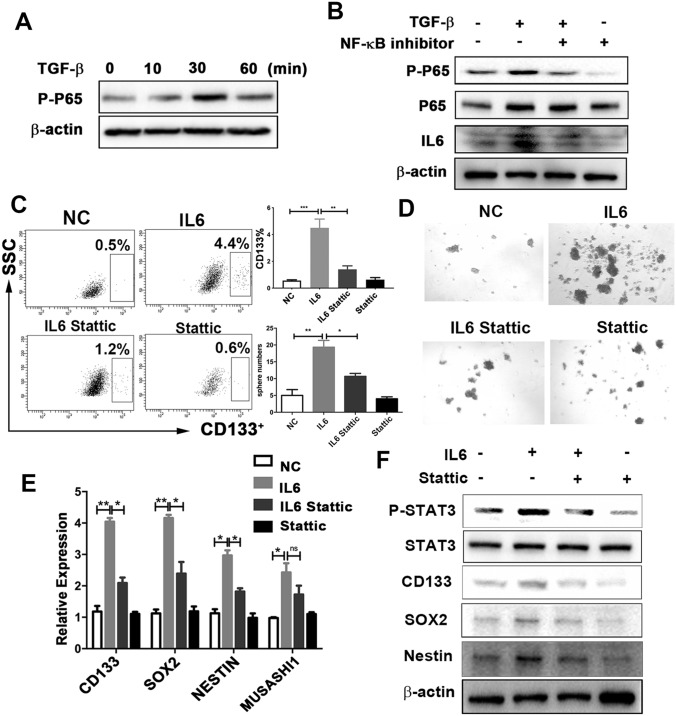
Fig. 5.
The STAT3 signaling pathway mediates IL6-induced GSC properties. a Expression of phospho-p65 under TGF-β treatment (10 ng/ml) at different times, assessed using western blotting analysis. b U251 cells were pretreated with NF-κB inhibitor (EVP4593) for 30 min, followed by TGF-β treatment (10 ng/ml) for 30 min and 24 h. The expression of phospho-p65 and IL6 was examined using western blotting analysis. c–f: U251 cells were pretreated with Stattic (10 umol/L, 30 min), followed by IL6 treatment (10 ng/ml, 24 h). c Cells were harvested to determine the proportion of CD133 cells. d Sphere numbers, determined using sphere-formation assay. The number of spheres per field was averaged from five random fields. e Expression of CD133, SOX2, NESTIN, and MUSASHI, analyzed using reverse-transcription PCR. f Expression of phospho-STAT3, STAT3, CD133, SOX2, and NESTIN, assessed using western blotting analysis. Results are based on three independent experiments. *P < 0.05; **P < 0.01; ***P < 0.001. NC normal control

