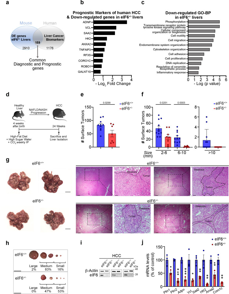
Fig. 5. eIF6 is required for NAFLD/NASH rapid progression into hepatocellular carcinoma.
a Comparison between prognostic and diagnostic markers of human hepatocellular carcinoma (HCC) and DE genes derived from RNA-Seq data of eIF6+/− livers: 159 common genes were found. b Representative downregulated genes in eIF6+/− livers identified as prognostic markers of human HCC. The histogram indicates the −Log2 fold change. c Gene Ontology (GO-BP) analysis identifies the most significantly downregulated pro-tumoral pathways in eIF6+/− livers. d Schematic overview of NAFLD/NASH/HCC progression mouse model. e, f Quantification of total surface tumors number (left) and size distribution (right) in 24 weeks eIF6+/+ (n = 11) and eIF6+/− (n = 8) livers. Two-tailed t test. g Gross appearance of livers and surface neoplastic nodules (left). Scale bars = 1 cm. H&E staining of liver sections of HFD/CCl4 mice at 24 weeks. Dashed black lines outline tumor areas surrounded by steatotic tissue. Scale bars = 500 μm (×4 magnification) and = 50 µm (×10 magnification). h Nodules size (as a percentage). Scale bars = 1 cm. i Representative western blotting for eIF6 on livers of HFD/CCl4 mice. j QT-PCR analysis of selected genes involved in lipid storage or associated with prognosis of HCC (n = 3 per genotype). Data are represented as means ± SD. Two-tailed t test. *p value ≤ 0.05, **p value ≤ 0.01, ***p value ≤ 0.001. NAFLD nonalcoholic fatty liver disease, NASH nonalcoholic steatohepatitis. Data are provided as a Source Data File.

