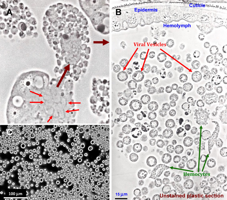
Figure 1.
Full cleavage of host cells to form viral vesicles in larvae of Trichoplusia ni infected with TnAV4. (A) Virus-directed cytopathology showing full cleavage of a fat body cell to form viral vesicles. The cell shown is greatly hypertrophied, with the small red arrows pointing to arrays of mitochondria that participate in the de novo formation of membranes that delimit nascent viral vesicles. The lower large maroon arrow points to nascent viral vesicles formed before the basement membrane ruptures, after which they spill into the hemolymph, indicated by the upper maroon arrow. (B) Viral vesicles circulating in the hemolymph where most transcription and progeny virion reproduction occurs. The vesicles with dense aggregates along the periphery of the vesicle membrane often have very few virions, and are thought to be spent vesicles, which have released most progeny virions into the hemolymph. (C) Phase contrast micrograph of a wet mount of vesicles in undiluted hemolymph of a larvae infected with TnAV4.

