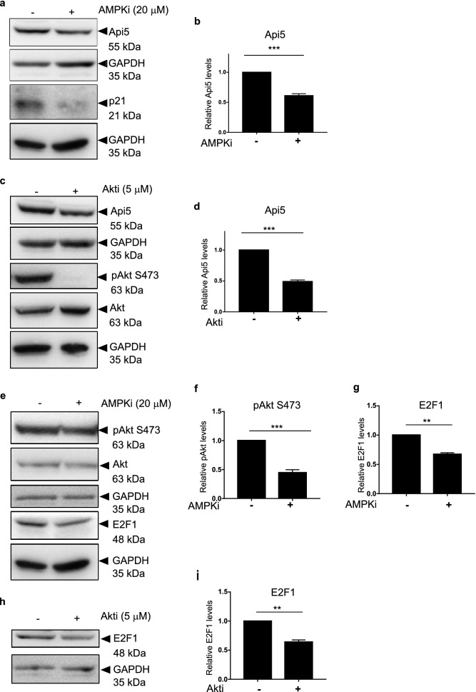
Figure 7.
AMPK and AKT regulate Api5 stability. MCF7 cells were treated with (a) 20 μM AMPKi, and (c) 5 μM AKTi for 4 h and 24 h, respectively. Immunoblotting analysis was performed using Api5 specific antibody to check for Api5 expression levels. GAPDH was used as loading control. (b,d) Quantification showing the fold change in Api5 levels after normalisation to GAPDH. (a) p21 and (c) pAkt S473 were used as positive control to confirm AMPK and Akt inhibition, respectively. (e) pAkt S473 activation upon AMPK inhibition. pAkt, Akt and GAPDH blots were cropped and the full blot is provided in Supplementary figure S12. (f) Quantification showing the fold change in pAkt S473 activation after normalisation to GAPDH. (e) E2F1 levels upon AMPK inhibition and (g) Quantification of E2F1 levels after normalising to GAPDH. (h,i) E2F1 protein levels were analysed following Akt inhibition using immunoblotting.

