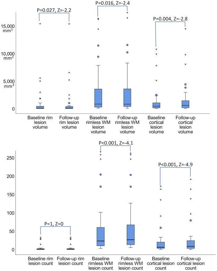
Figure 3.
Boxplots summarizing the longitudinal changes of multiple sclerosis lesions in 46 patients. The cortical and rim lesion volumes increased over time while the rimless white matter volume decreased (A) despite the new white matter lesion formation (B) in multiple sclerosis patients. P and Z-statistic values by Wilcoxon signed rank test (related samples, two-tailed).

