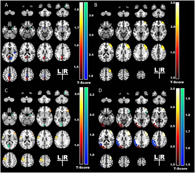
Figure 2.
The heatmap represents the t statistics* of the univariate analysis and displays the structures that were identified by the model as outcome predictors. (A) Failure of postoperative seizure control for right-sided temporal lobe surgeries associated with smaller volumes of the transverse temporal, pericalcarine and entorhinal cortex in the right (ipsilateral) hemisphere, and with larger volumes of medial parietal in the left (contralateral) hemisphere. Seizure recurrence also associated with asymmetry of nucleus accumbens and paracentral regions, with the left side (contralateral hemisphere) smaller than the right. (B) Failure of postoperative seizure control for left-sided temporal lobe surgeries associated with smaller volumes of the right middle frontal region in the right (contralateral) hemisphere, and with asymmetry of middle frontal gyri, with the right side (contralateral hemisphere) smaller than the left. (C) Worse outcomes (Engel II–IV) for right-sided temporal lobe surgeries associated with smaller volumes of the entorhinal cortex in the right (ipsilateral) hemisphere, and with smaller volumes of the pericalcarine cortex and with larger volumes of the primary motor cortex in the left (contralateral) hemisphere. (D) Worse outcomes (Engel II–IV) for left-sided temporal lobe surgeries associated with smaller volumes of the pars orbitalis in the right (contralateral) hemisphere and asymmetry of the occipital lobe, and inferior parietal region with the left side (ipsilateral) hemisphere smaller than the right and asymmetry of middle frontal region with the right side (contralateral hemisphere) smaller than the left. * t-statistics represented in colour bars: cool colours negative t-values and hot colours positive t values.

