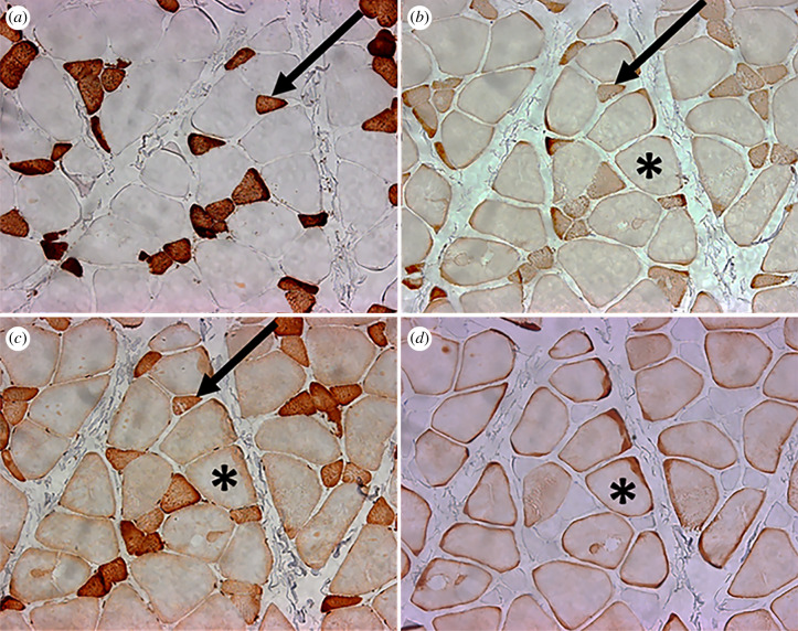
Figure 3.
20× images of the same muscle fascicle in the anterior superficial masseter of M. fascicularis. (a) NOQ7.5.4D (MHC-1); (b) MYH6 (MHC α-cardiac); (c) MY32 (MHC-2); (d) (MHC-M). Note the abundance of hybrid fibres. Arrows point to the same cell co-staining with intermediate or dark intensity for MHC-1, MHC α-cardiac and MHC-2 (the slow + 2 hybrid). Asterisks indicate the same cell co-staining with light or intermediate intensity for MHC α-cardiac, MHC-2 and MHC-M (the fast + α-cardiac hybrid). Note the counter-staining between (a) cells that express MHC-1 and (d) those that express MHC-M.

