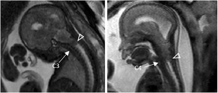
FIGURE 7.

Left: Fetal magnetic resonance (MR) image of a Myelocele patient at an estimated gestational age of 27 weeks showing a vermian displacement (arrowhead) at C3. Right: Fetal MR image of a Myelocele patient at an estimated gestational age of 22 weeks showing vermian displacement (arrowhead) at C4. The exact level of displacement was determined by counting the T2‐weighted hyperintense intervertebral discs (arrow)
