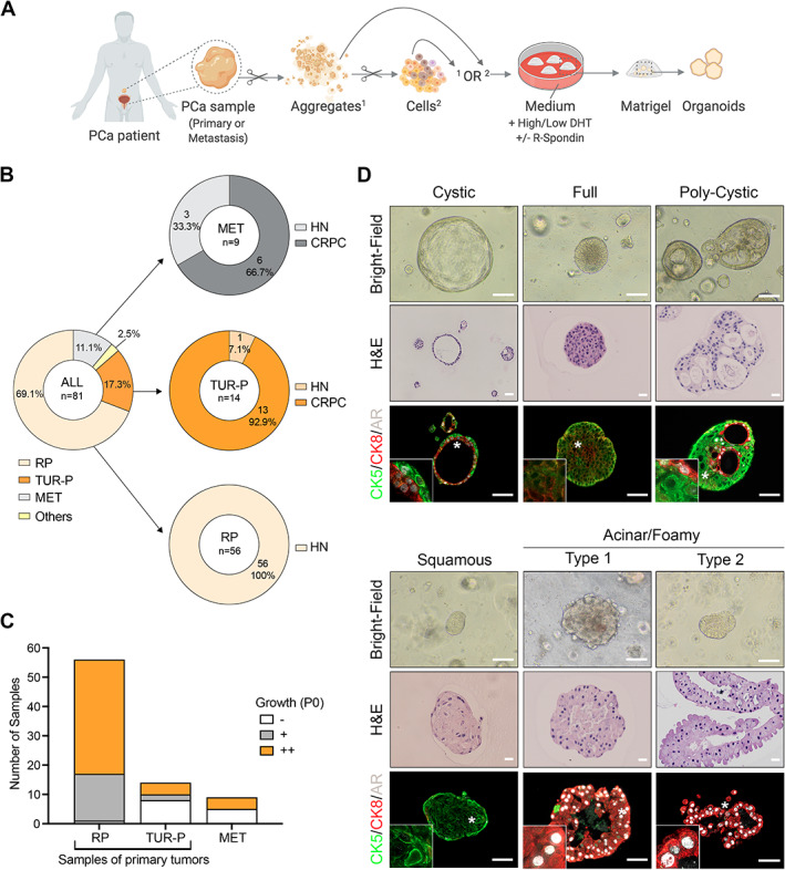
Figure 1.

Generation of organoids derived from PCa patients. (A) Schematic diagram depicting the experimental strategy and the distinct conditions used to establish organoids derived from primary PCa or PCa metastasis. Aggregates (1) or cell suspensions (2) are seeded in Matrigel domes immersed in organoid medium. Scissors symbols indicate mechanical or enzymatic digestion. Created with BioRender.com. (B) Summary of sample types used to establish organoid cultures (n = 81). RP, radical prostatectomy; TUR‐P, transurethral resection of the prostate; MET, metastasis. ‘Others’ corresponds to a primary PCa resection obtained from an autopsy of a man with metastatic CRPC and one local progression of primary PCa obtained from a bladder resection. HN, hormone‐naïve; CRPC, castration‐resistant PCa. (C) Growth efficiency for each sample category at passage 0 (P0). Group ‘–’: no significant organoid growth; group ‘+’: between one and five organoids per well; group ‘++’: more than five organoids per well. (D) Bright‐field images, corresponding H&E staining, and immunofluorescence analysis for the indicated antibodies in selected organoids representative of five major morpho‐histological subtypes. Insets show higher magnification of a representative region indicated by a white asterisk on the original image. Scale bars: 100 μm for bright‐field images; 20 μm for H&E images; 50 μm for immunofluorescence images.
