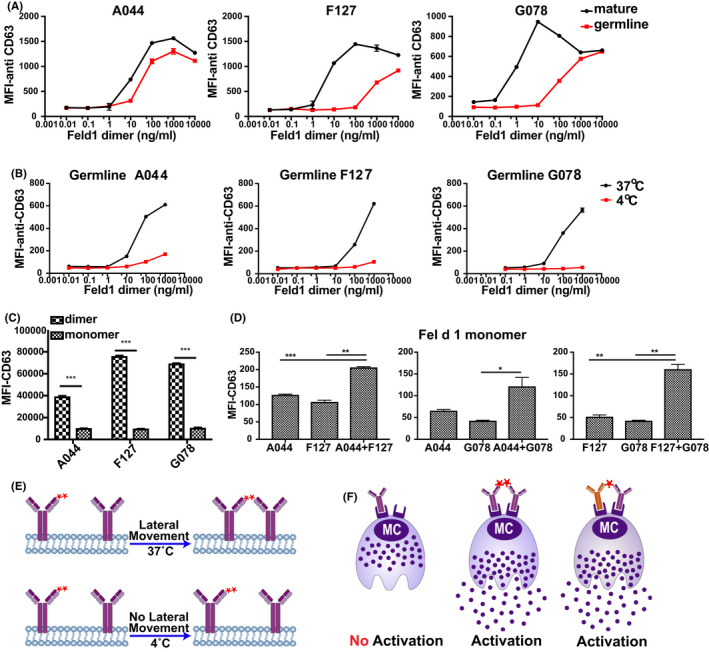
FIGURE 3.

Activation of BMMCs after Fel d 1 binding to mature and germline IgE antibodies in vitro. Mean fluorescent intensities of anti‐CD63‐FITC were analysed after BMMCs were incubated with IgE antibodies overnight and followed by 10000, 1000, 100, 10, 1, 0.1 and 0.01 ng/ml Fel d 1 dimer (A); the same measurement was performed to determine the activation of BMMCs after germline IgE incubation at 4°C and 37°C (B); (C) illustrates the activation of BMMCs by Fel d 1 dimer and monomer (1000 ng/ml) after mature and germline IgE antibodies incubation overnight; activation of BMMCs upon Fel d 1 monomer (1000 ng/ml) binding to different germline IgE antibody combinations overnight was shown in (D); (E) illustrates the binding pattern of Fel d 1 dimer to germline IgE antibodies with and without lateral movements; (F) summarizes different Fel d 1 molecules binding to germline IgE antibodies on cell surface and the activation status
