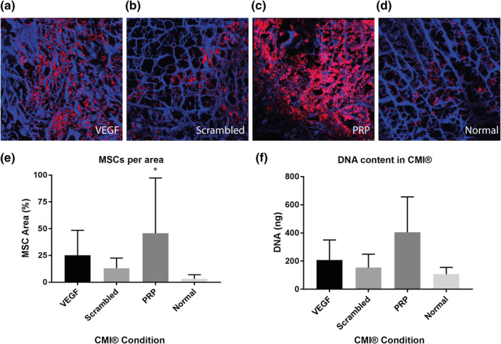
FIGURE 6.

Cell migration of mesenchymal stromal cells (MSCs) into the Collagen Meniscus Implant (CMI®) (n = 6). The CMI® is stained with DAPI (blue) and MSCs with DiI (red) (A–D). The figure shows CMI® with EDC/NHS, VEGF peptide and VEGF (A), and CMI® with EDC/NHS, a scrambled VEGF binding peptide and VEGF (B), CMI® coated with PRP (C) and CMI® (D). E corresponds with A–D, and shows percentage of meniscus cells per CMI® area. F shows DNA quantification in the whole constructs. *p < 0.05
