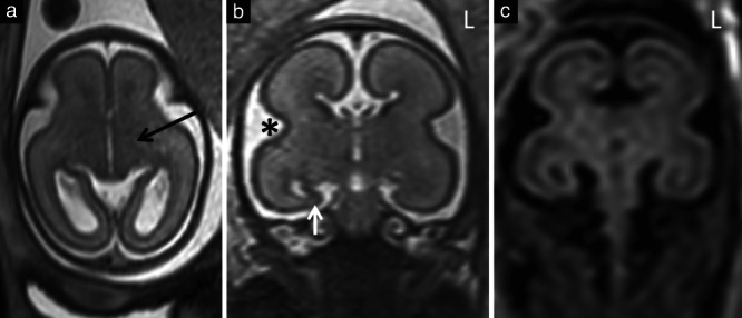
Figure 3.

Example brain magnetic resonance images (MRI) in a fetus with isolated corpus callosal agenesis at 23 weeks' gestation that had a low MRI score. (a,b) T2‐weighted single‐shot fast‐spin echo images in axial (a) and coronal (b) planes. (c) T2‐weighted FLAIR image in coronal plane. There was no ventriculomegaly and normal basal ganglia (internal capsule (black arrow)) (a), normal opercularization () (b) and normal lamination, which was better identified on T2‐weighted FLAIR imaging (c). Features included unilateral mild hippocampal malrotation on the right (white arrow) (b), scoring 1 point, and inverted temporal lobe symmetry (b,c), with a ‘squarer’ temporal lobe in the coronal plane on the left (L), scoring 1 point.
