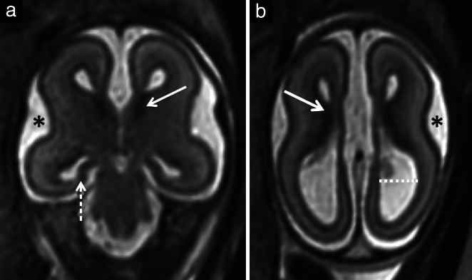
Figure 4.

Example brain magnetic resonance images (MRI) in a fetus with isolated corpus callosal agenesis at 22 weeks' gestation that had a high MRI score. Coronal (a) and axial (b) T2‐weighted single shot fast‐spin echo images. There was delayed opercularization () (a,b), scoring 1 point, severely malrotated hippocampi that were almost completely flat (dashed arrow) (a), scoring 3 points, abnormal lamination with abnormally thick germinal matrix (solid arrows) (a,b), scoring 2 points, and moderate ventriculomegaly (dotted line) (b), scoring 1 point. (b) Unlike in Figure 3, lamination could be visualized adequately on T2‐weighted image.
