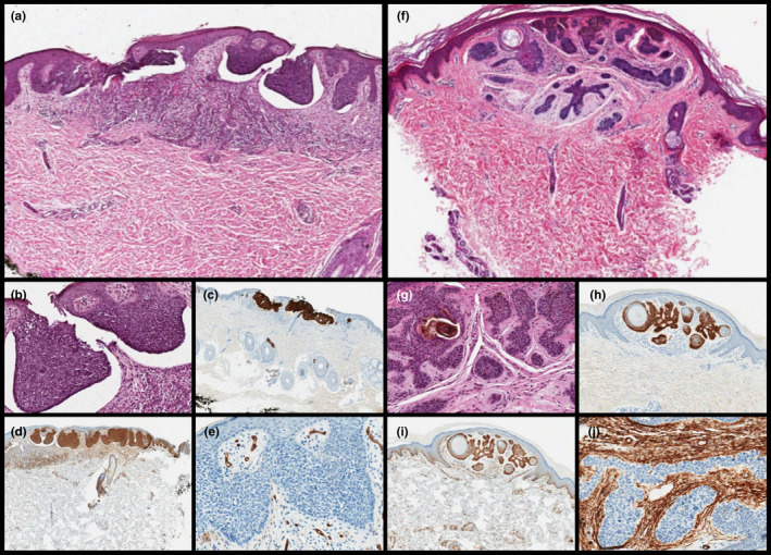
FIGURE 2.

(a–b) Scanning and high magnification shows basal cell carcinoma (BCC) with superficial proliferation of basaloid cells, peripheral palisading and clefts within between the epithelial component and the typical fibrous and mucinous stroma (a, H&E, original magnification ×60; b, original magnification ×200). The lesion was diffusely positivity for (c) Ber‐EP4 (original magnification ×60) and (d) BCL2 (original magnification ×60), (e) with stroma negative for CD34 (original magnification ×200). (f–g) Scanning magnification showed BFH with the so‐called "inverted candlestick" shape, radially emerging from the follicular axis and replacing the affected follicular unit with no destruction of the interfollicular dermis. Note the infundibular cysts with lamellar keratin were inside the basaloid epithelial cords (f, H&E, original magnification ×60; g, H&E, original magnification ×200). The lesion was diffusely positivity for (h) Ber‐EP4 (original magnification ×60) and moderately (with the typical peripheral staining) for (i) BCL2 (original magnification ×60), (j) with stroma negative for CD34 (original magnification ×200)
