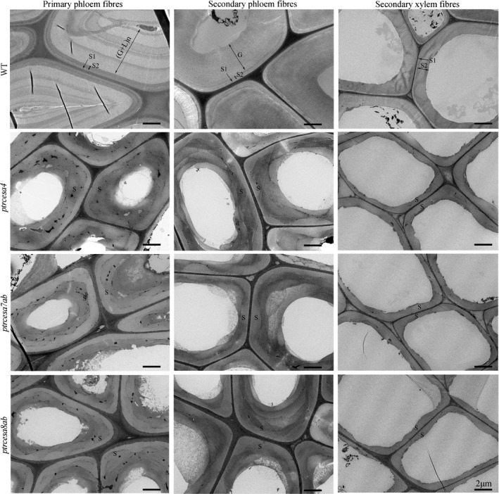
Fig. 5.

Transmission electron microscopy analysis of the phloem and wood fibre wall structures in Populus trichocarpa ptrcesa4, ptrcesa7a/b and ptrcesa8a/b mutants (CesA, cellulose synthase). Transmission electron microscopy (TEM) images from the basal stem phloem and xylem fibres in 6‐month‐old wild‐type (WT) and ptrcesa mutant trees. The WT primary and secondary phloem fibres showed S1 + S2 + n(G + L) and S1 + S2 + G wall structures, respectively. L, lignified layer; G, gelatinous (G)‐layer; n, number of repetitions of the G and L; S, S‐layer of SCW with S1, S2 and S3. Bars, 2 μm.
