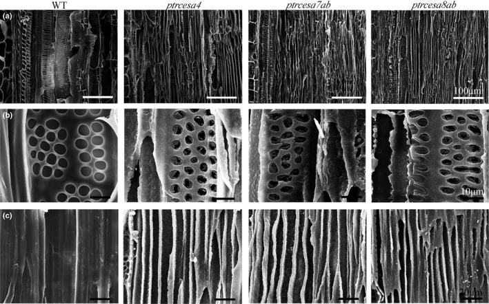
Fig. 6.

Scanning electron microscopic images of xylem fibres and vessels in longitudinal sections of basal stems from Populus trichocarpa wild‐type (WT) and ptrcesa4, 7a /b and 8a /b mutants (CesA, cellulose synthase). (a‐c) Primary and secondary xylem (a), the pitted pattern vessels of secondary xylem (b) and the fibres of secondary xylem (c). Bars: (a) 100μm; (b) 10 μm; (c) 20 μm.
