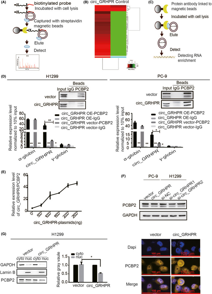
FIGURE 3.

Circ_GRHPR interacts with the RNA‐binding protein PCBP2 and regulates its subcellular localisation. (A) The schematic diagram provides a summary of the RNA pull‐down procedure. (B) The cluster analysis showed 58 candidate proteins that might interact with circ_GRHPR. (Red and green represent up‐ and downregulation, respectively). (C) The schematic diagram shows the RIP procedure. (D) circ_GRHPR molecules enriched by PCBP2 protein and IgG protein were detected by RIP assay in H1299 and PC‐9 cells. α‐globin is a positive control that has been shown to interact with PCBP2; γ‐globin was the negative control. Western blotting shows the reliability of the RIP experimental system data. (E) The relationship between the amount of circ_GRHPR/PCBP2 complex and circ_GRHPR was observed by the RIP experimental system. With an increase in the circ_GRHPR plasmid, the expression levels of circ_GRHPR increased and then the number of circ_GRHPR enriched by PCBP2 increased (F) PCBP2 expression in PC‐9 and H1299 cells was detected by western blotting after transfecting the circ_GRHPR overexpression plasmid and siRNA, respectively. (G) PCBP2 expression levels in the nucleus and cytoplasm of circ_GRHPR‐OE and NC cells were detected in H1299 cells. The grey value of their protein bands is shown on the right. (H) The location of PCBP2 protein was detected in circ_GRHPR‐OE and NC cells of H1299 by FISH. Data are presented as the mean ± SD, p < 0.05 was considered statistically significant (*p < 0.05/**p < 0.01/***p < 0.001)
