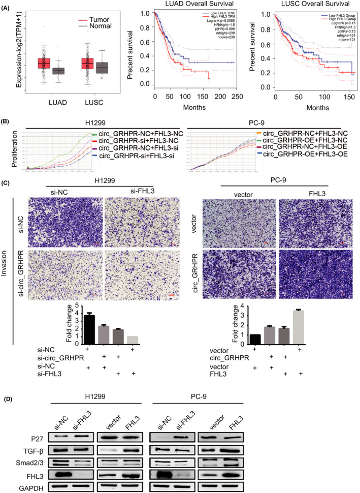
FIGURE 5.

FHL3 promotes proliferation and invasion of non‐small cell lung cancer (NSCLC) cells. (A) Expression levels of FHL3 and overall survival of the FHL3 genes were validated in LUAD, LUSC, and normal tissues with GEPIA2. |log2FC| > 1 and p‐value <0.01 were considered statistically significant. Tumour and normal tissues are shown in red and grey, respectively. (B) The proliferation status of FHL3 was determined by the RTCA assay in H1299 and PC‐9 cells. (C) The invasion status of FHL3 in H1299 and PC‐9 cells was determined by a transwell assay. The number of cells was quantified in five fields under a 10× microscope. (D) P27, TGF‐β, and Smad2/3 were downstream target genes of FHL3 in NSCLC cells. Data are presented as the mean ± SD, p < 0.05 was considered statistically significant (*p < 0.05/**p < 0.01/***p < 0.001)
