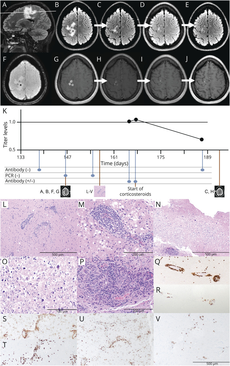
Figure. Serial MRIs, Time Course, and Histopathology.
(A, B, F, G) Initial brain MRI (5 months after presumed COVID-19 infection). The white line in A depicts the level of the axial sections in B–D. Sagittal (A) and axial (B) T2/FLAIR, susceptibility-weighted (F), and gadolinium-enhanced T1-weighted (G) sequences show multiple peripherally irregularly enhancing ovoid lesions within the right frontoparietal white matter with surrounding T2/FLAIR hyperintensity suggestive of vasogenic edema. (C–E, H–J) Repeat MRI at 1 (C, H), 3 (D, I), and 6 (E, J) months after the initiation of steroid treatment. Axial T2/FLAIR-weighted (C–E) and contrast-enhanced T1-weighted (H–J) sequences show a decreased size of T2/FLAIR hyperintensities and decreased contrast enhancement. (K) Time course and results of antinucleocapsid SARS-CoV-2 antibody testing. Time is shown as days since COVID-19 infection. The upper graph depicts the time course of available Roche titer levels (1.03, 1.05, and 0.7; the assay's cutoff of 1.0 is marked as a solid black line). Time point of initial and repeat MRI, biopsy, and initiation of corticosteroids is shown below the timeline. (L–V) Stereotactic biopsy of the right frontal lesion. (L–P) H&E-stained sections show edematous and gliotic white matter with extensive lymphoplasmacytic perivascular inflammation, as well as infiltration of vessel walls by lymphocytes, consistent with lymphocytic vasculitis (L: ×100; M: ×200). Multiple foci of eosinophilic coagulative necrosis with extensive dystrophic calcification were also noted (N: ×100; O: ×400). Focal endothelial swelling and hypertrophy, as well as vascular proliferation were present (P: ×200). (Q–V) Immunohistochemistry showed the perivascular and intravascular lymphocytes to be predominantly CD3-positive T cells (Q: CD3 IHC, ×100) with a small subset of CD20-positive B cells (R: CD20 IHC, ×100). Numerous perivascular CD4-positive T lymphocytes (S: CD4 IHC, ×100), many perivascular and parenchymal CD8-positive T lymphocytes (T: CD8 IHC, ×100), abundant perivascular macrophages and parenchymal activated microglia (U: CD68 IHC, ×100), and scattered perivascular plasma cells (V: CD138 IHC, ×100) were observed. Special stains for bacterial and fungal organisms including Gram, AFB, GMS, and HSV1/2 stains were negative (not shown) as well as tissue cultures. No viral inclusions were seen. AFB = acid-fast bacteria; COVID-19 = coronavirus disease 2019; FLAIR = fluid-attenuated inversion recovery; GMS = Grocott's methenamine silver; H&E = hematoxylin and eosin; HSV = herpes simplex virus; IHC = immunohistochemistry.

