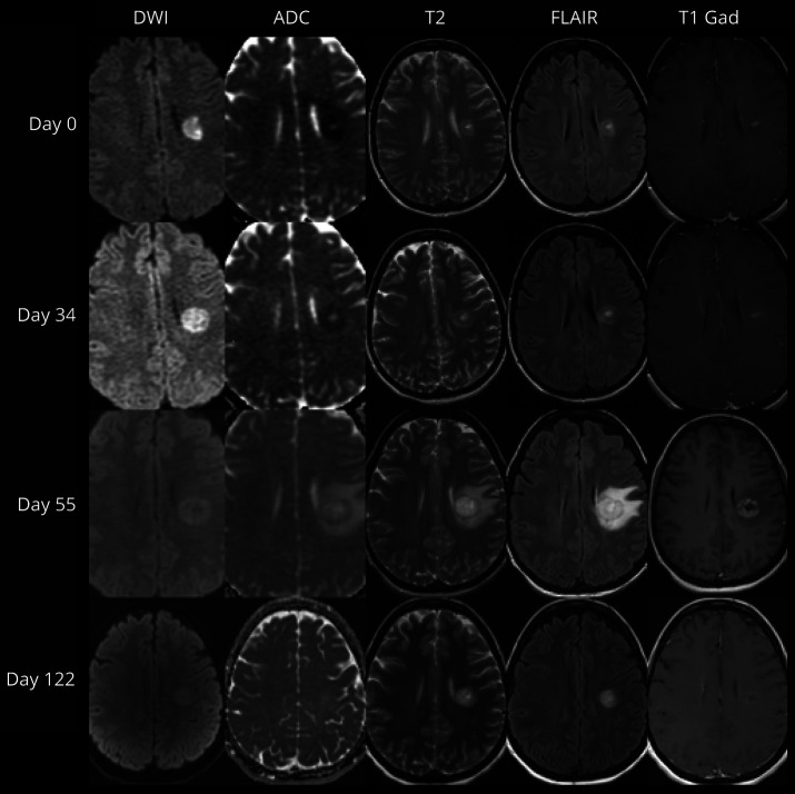
Figure 2. Temporal Evolution of a Baló Concentric Sclerosis.
Diffusion-weighted imaging (DWI), apparent diffusion coefficient (ADC), T2-weighted (T2), fluid-attenuated inversion recovery (FLAIR), and postgadolinium T1-weighted (T1 Gad) MRI demonstrating evolution of a single lesion with initial peripheral diffusion restriction associated with central enhancement progressing to develop multiple concentric rings, associated with peripheral enhancement, followed by resolution of enhancement and ADC changes with T2-weighted abnormalities being persistently visible at 122 days from the initial MRI.

