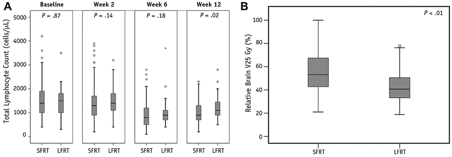
Fig. 1.

(A) Box and whisker plot illustrating median and quartile distribution of total lymphocyte counts for standard-field radiation therapy (SFRT) and limited-field radiation therapy (LFRT) patients at baseline, week 2, week 6, and week 12. (B) Box and whisker plot illustrating median and quartile distribution of brain V25 Gy (brain volume receiving 25 Gy) for standard-field radiation therapy and limited-field radiation therapy patients.
