Figure 5.

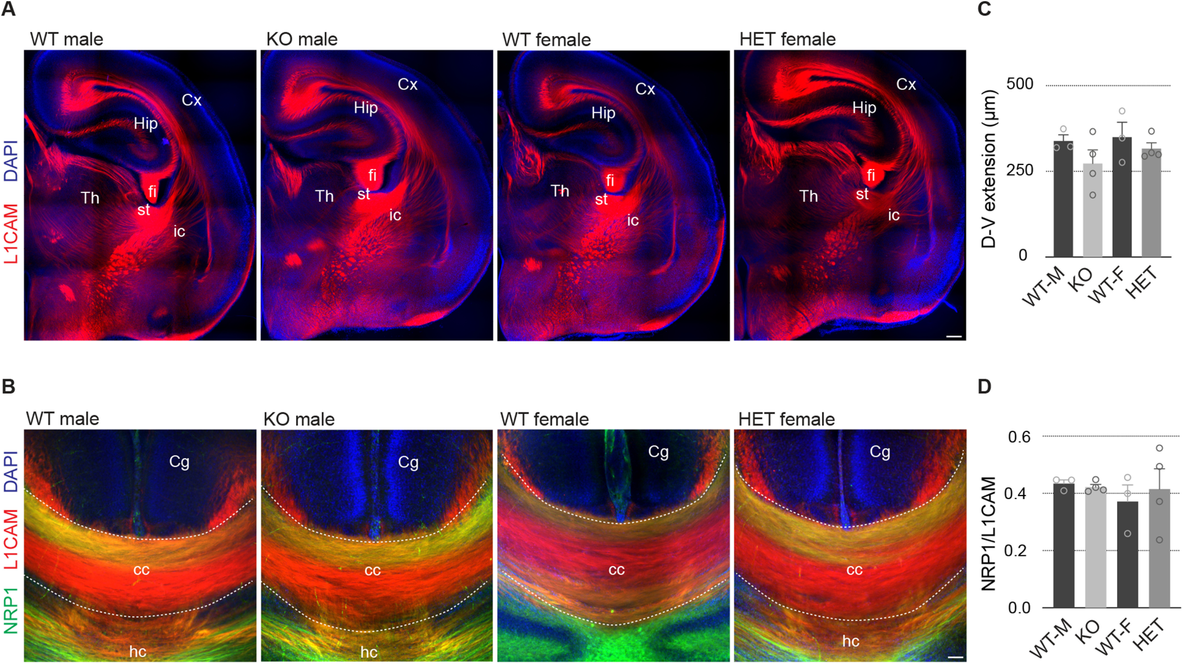
No major anomalies in the main axonal tracts in Pcdh19 mouse mutants. A, Confocal micrographs of P0–P1 mouse hemispheres stained with anti-L1CAM (red). Nuclei were counterstained with DAPI (blue). B, Confocal micrographs of the corpus callosum of P0–P1 mice stained with anti-L1CAM (red) and anti-Neuropilin-1 (green), and counterstained with DAPI (blue). C, Quantification of the dorsoventral extension of the corpus callosum in WT and mutant animals, separated by sex. D, Quantification of the dorsal restriction of Neuropilin-1+ axons in WT and mutant animals, separated by sex. All results are indicated as the mean ± SEM. Two images per brain, obtained from four animals originating from three different litters, were analyzed for each condition. Cx, Cortex; Hip, hippocampus; Th, thalamus, fi, fimbria; st, striatum; ic, internal capsule; Cg, cingulate cortex; cc, corpus callosum; hc, hippocampal commissure. Scale bars: A, 200 μm; B, 50 μm.
