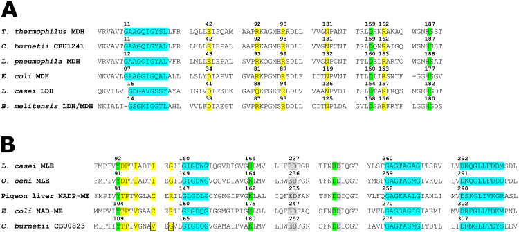
Fig 2. Key residues of CBU1241 and CBU0823.
Protein sequence alignment sections containing key residues of (A) CBU1241 compared with MDHs and LDHs and (B) CBU0823 compared with MEs and MLEs. The NAD(P)-binding motifs (blue), substrate binding residues (yellow), catalytic activity residues (green), and metal binding residues (grey) are highlighted, and the boxes indicate unusual residues present within CBU0823 versus other bacterial MEs. Numbering refers to the residue position within the respective protein.

